
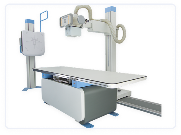
X-Ray Machine
An X-ray machine is a medical imaging device that uses X-rays (a form of electromagnetic radiation) to create detailed images of the inside of the body. It is primarily used to examine bones, tissues, and organs, helping healthcare professionals diagnose and monitor various medical conditions, such as fractures, infections, tumors, and diseases affecting internal organs.
Key Features of an X-Ray Machine
X-ray Tube
The X-ray tube is the core component of the machine that generates the X-rays. It contains a cathode (negative electrode) and anode (positive electrode) that work together to produce the X-rays when electricity flows through the tube.
Detector
The detector captures the X-rays that pass through the body and converts them into an image.
Collimator
The collimator is a device that narrows the X-ray beam to a specific area of interest. This reduces unnecessary exposure to radiation in other parts of the body and ensures more focused imaging.
Control Panel
The control panel is used by the technician to set the exposure parameters, including radiation dose, exposure time, and X-ray energy (kVp), depending on the area being imaged and the patient's size.
High-Voltage Generator
This component provides the high voltage needed to accelerate electrons from the cathode to the anode, generating X-rays.
Digital Imaging
Digital X-ray systems capture images on a digital sensor rather than film. These systems offer benefits such as instant image viewing, the ability to adjust contrast, and easy digital storage for long-term records.
Fluoroscopy
Some X-ray machines include fluoroscopy, which provides real-time, continuous imaging. This is particularly useful in procedures like guiding catheter placements, barium studies, and real-time monitoring of a patient's anatomy during certain interventions.
Patient Positioning Equipment
X-ray machines are often equipped with special positioning tables or arms to position the patient correctly during the imaging process.
Radiation Safety Features
Lead shielding or lead aprons are commonly used in conjunction with the machine to protect both the patient and the technician from unnecessary radiation exposure.
Mobile and Stationary Units
Mobile X-ray machines are portable, allowing for X-ray imaging in emergency rooms, intensive care units (ICUs), or patient rooms without needing to transport the patient to a radiology department.
Image Storage and Transmission (PACS)
Picture Archiving and Communication System (PACS) allows for the digital storage, retrieval, and sharing of X-ray images across different devices and locations.
Various industries
Request More Information
Contact Us to learn more about our offerings or Request a Quote to get started today!
Contact Us

